Spatial Technologies

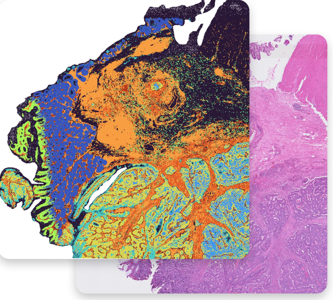
Spatial technologies are revolutionizing biology by offering a whole new perspective on tissues. Unlike traditional methods that analyze cells in isolation, these tools capture the gene activity and protein expression of cells within their actual tissue location. This is like looking at a painting instead of just a collection of pigments - you see how different elements work together. This deeper understanding allows researchers to untangle complex biological processes, identify new cell types, and pinpoint how diseases develop within tissues.

The following spatial technologies are available to researchers at The University of Sheffield. Talk to us and we will advise on which will best suit your experiment.

Visium Spatial Xenium in-situ
10X CytAssist for visium and Visium HD (both FFPE)
  • Sequencing-based
  • Focus: Broad gene expression analysis across a tissue section
  • Data capture:Whole transcriptome (all RNA molecules)
  • Strengths:
    • Unbiased discovery of gene expression patterns
    • Provides a general picture of gene activity across many cell types
    • Allows for H&E or immunofluorescence staining upfront for initial screening
  • Weaknesses
    • Lower per-gene sensitivity compared to Xenium
    • Might miss rarer transcripts due to broader analysis
  • 10X Xenium Analyser - Available via the School of Clinical Dentistry
  • Imaging-based
  • Focus: Targeted, high-resolution analysis of specific genes
  • Data capture: Pre-defined set of genes
  • Strengths:
    • High per-gene sensitivity
    • Offers detailed information on chosen genes and their spatial distribution
    • Allows for H&E or immunofluorescence staining at the end of the workflow
  • Weaknesses:
    • Requires prior knowledge of genes of interest
    • Lacks the broad discovery potential of Visium

  • For queries relating to collaborating with the Genomics teams at The University of Sheffield please email: genomics-group@sheffield.ac.uk